|
Consultul de specialitate in cadrul Ambulatoriului de specialitate al CLINICII SOMEȘAN este realizat de către Dr. TODINCA ADRIANA si Dr. POP MUNTEAN LAVINIA
SÂNGELE
Sângele este constituit dintr-o componentă lichidă, plasma sangvină şi o componentă solidă, reprezentată de elementele figurate. Sângele îndeplineşte două funcţii majore: transportul in organism al: oxigenului si dioxidului de carbon, moleculelor nutritive, ioni (Na+, Ca2+, HCO3-, etc), produşilor de excreţie (uree, bilirubina, etc), hormoni si apărarea organismului, proces în care sunt implicate toate celulele albe ale sângelui.

Plasma sangvină conţine apa (în proporţie de peste 90%) în care sunt dizolvate substanţe anorganice (in special ioni) si substante organice (proteine, substante nutritive, produsi de metabolism, hormoni, etc). Plasma sangvină din care au fost îndepărtate proteinele de coagulare reprezintă serul. Elementele figurate se pot clasifica în: celule roşii sau hematii/eritrocite, celule albe (leucocite) şi plachete sangvine sau trombocite.
HEMATIILE
Sunt cele mai numeroase elemente figurate, în jur de 4.8 x 10⁶/mm³ la femei si 5.5 x 10⁶/mm³ la bărbaţi. Aceste valori pot varia în funcţie de factori precum starea de sănătate, vârsta, altitudine (peruvienii care traiesc la altitudini de peste 5.400 m pot avea pana la 8.3 x 10⁶ hematii/mm³. Sunt celule anucleate, cu forma de disc biconcav. RBC se dezvolta in hematopoieza din celule progenitoare eritroide. Pe parcursul procesului de maturare, celulele produc hemoglobina până ce aceasta ajunge să reprezite 90% din greutatea uscata a celulei. Celulele se maturează în apropierea unui macrofag care va ingera nucleul eritrocitului.

Celulele roşii sunt responsabile cu transportul oxigenului si dioxidului de carbon in organism. Transportul oxigenului: molecula de hemoglobina este constituita din 4 polipeptide (doua lanturi alfa si doua lanturi beta) fiecare dintre acestea fiind ataşată la o grupare prostetica, denumita grupare hem. Fiecare grupare hem contine un atom de fier care va lega o molecula de oxigen si se va forma oxihemoglobina. Reactia este reversibila.
Hemoglobina este principalul component al hematiei. Este o cromoproteină, alcătuită din două componentei o proteină, numită globină şi o grupare neproteică numită hem. Globina este constituită prin asocierea a patru lanţuri polipeptidice.
De fiecare lanţ polipeptidic se leagă câte o moleculă de hem. Datorită prezenţei Fe în molecula sa, hemul poate lega labil oxigenul .Reacţia de fixare a 02 ia Hb nu este o oxidare propriu-zisă (deoarece ea nu duce la creşterea valenţei Fe) ci o reacţie de oxigenare, de legare reversibilă a unei molecule de oxigen la fierul bivalent, în urma acestei reacţii rezultă oxihemoglobină (HbO2) care reprezintă forma principală de transport a O2 prin sânge. Atunci când este saturată (oxigenata) complet, o moleculă de HB poate transporta 4 molecule de 02. O singură hematie conţine cam 300 000 000 molecule Hb. Exprimată în grame, Hb reprezintă 16 g la o sută de ml sânge. Fiecare gram de Hb poate transporta 1,34 ml 02, deci fiecare sută de ml sânge transportă 20 ml 02. În lipsa Hb, capacitatea de transport a sângelui pentru oxigen scade mult ; 100 ml plasmă transportă doar 0.2 ml 02.
În afară de forma oxigenată şi cea redusă Hb poate da compuşi stabili cu oxidul de carbon (CO Hb) denumiţi carboxihemoglobină iar sub acţiunea oxidanţilor apare derivatul de Hb cu Fe trivalent, denumit methemoglobină. Aceştia sunt derivaţi patologici ai Hb; ei nu mai îndeplinesc funcţia de transport şi în cazul creşterii concentraţiei lor în sânge peste anumite limite se produce insuficienta oxigenare a ţesutului (asfixie).

Hemoglobina se poate combina şi cu dioxidul de carbon (Hb C02), compus numit carbohemoglobina sau carbamatul de hemoglobina. Acesta este un compus fiziologic, ce nu afectează funcţia de transport a 02. HbC02 reprezintă şi una din formele de transport ale C02 de la ţesuturi la plămâni.
LEUCOCITELE (CELULELE ALBE)
Sunt mult mai putin numeroase decât hematiile (4000-8000 /mm3). Sunt celule nucleate care participa la apararea imuna a organismului. In functie de prezenta sau absenta granulatiilor din citoplasma se clasifica in celule fara granulatii (limfocite si monocite) si granulocite (celule cu granulatii): neutrofile, eozinofile si bazofile.
Forma leucocitelor nu este aceeaşi. Ele nu reprezintă o populaţie celulară omogenă. Există mai multe tipuri, care diferă între ele atât ca origine şi morfologie cât şi în privinţa rolului în organism. Exprimarea lor procentuală se numeşte formulă leucocitară. În cadrul acestei formule, deosebim leucocite cu nucleu unic — mononucleare şi cu nucleu fragmentat, polilobat - polinucleare.

Mononuclearele reprezintă 32% iar polinuclearele 68% din leucocite. În grupa mononuclearelor se cuprind: limfocitele, care reprezintă 25% şi monocitele, 7%.
Dimensiunile leucocitelor variază între 6-8 µ pentru limfocitul mic şi 20 µ, în diametru pentru monocite şi neutrofile. Leucocitele prezintă o structură celulară completă. Au o membrană cu o plasticitate remarcabilă. Datorită ei leucocitele întind prelungiri citoplasmatice (pseudopode), cu ajutorul cărora devin mobile, se pot deplasa în afara vaselor capilare (diapedeză) şi pot îngloba microbi (microfagocitoză) sau resturi celulare (macrofagocitoză). Granulaţiile polinuclearelor sunt mici saci şi vezicule pline cu enzime hidrolitice (lizozomi) care participă la digestia corpului fagocitat. Tot în familia leucocitelor se includ şi plasmocitele, celulu provenite din limfocite, specializate în producţia de anticorpi.
Rolul leucocitelor este complex şi diferit, după tipul lor. Principala funcţie a leucocitelor constă în participarea acestora la reacţia de apărare a organismului.
LIMFOCITE
Toate limfocitele sunt produse în măduva osoasă (un organ limfoid primar). Dacă devin imunocompetente în măduva osoasă se numesc limfocite B (sintetizeaza anticorpi si limfokine) iar dacă devin imunocompetente în timus (un alt organ limfoid primar), se numesc limfocite T (sintetizează doar limfokine). Exista mai multe tipuri de limfocite T, cele mai comune fiind:
- limfocitele T inflamatorii care recrutează macrofage si neutrofile la situsul unei infectii sau al unei leziuni tisulare;
- limfocite T citotoxice (CTL) care ucid celule infectate cu virusuri si (probabil) celule tumorale
- celule T helper - stimuleaza producerea de anticorpi de catre limfocitele B.

MONOCITE
Monocitele circulă prin sângele periferic înainte de a emigra în ţesuturi unde se transformă în macrofage. In functie de organul în care sunt localizate au denumiri specifice. Astfel macrofagele din ficat se numesc celule Kupfer, în creier se numesc celule microgliale, în os - osteoclaste, etc. Macrofagele sunt celule mari, fagocitare care înglobează materiale straine organismului sau celule si fragmente de celule ale organismului.
NEUTROFILE
Neutrofilele sunt elemente sangvine care răspund la semnale chemotactice si părăsesc capilarele printr-un proces complex care implică marginaţia celulelor (apropierea de endoteliul vaselor sangvine), ataşarea la peretele vasului şi ieşirea din capilar prin spatiul dintre celulele endoteliale (proces denumit extravazare sau diapedeza). Migrarea este determinată de mai multi factori: substanţe produse de microorganisme, semnale emise de celule participante la procesul inflamator, etc. Neutrofilele ţin sub control populaţiile de bacterii comensale existente în mod normal în organismul uman în colon, cavitatea bucală sau la nivelul gâtului. In cazuri patologice, când numărul de neutrofile scade (radiatii, chemoterapie, stress), aceste bacterii scapă de sub control, proliferează excesiv şi apar infectiile oportuniste.
EOZINOFILE
Numărul de eozinofile este în mod normal cuprins intre 0 si 450/mm3. Numărul lor creşte în anumite boli, în special in cazul parazitozelor, mai ales in cazul parazitilor mari. Granulele eozinofilelor contin substante citotoxice, care sunt eliberate asupra parazitului. Printre substantele din granule se numără proteina bazică majoră (MBP - major basic protein), proteine cationice, peroxidaza, arilsulfataza B, fosfolipaza D si histaminaza. Acest amestec este capabil sa distruga membranele parazitului.
BAZOFILE
Bazofilele sunt celule nefagocitare care, atunci când sunt activate eliberează numeroşi compuşi din granulele bazofile din citoplasma lor. Joacă un rol major în răspunsurile alergice, în special în cazul reactiilor hipersenzitive de tip I. Bazofilele sunt implicate în răspunsul anafilactic. Anafilaxia este o reacţie de hipersensibilitate specifică, care apare la a doua expunere la acelesi antigen. Forma cea mai gravă este şocul anafilactic. O bună definiţie clinică a reacţiei anafilactice tine cont de prezenţa a două manifestări severe: dificultatea respiratorie (prin edem laringian sau criza de astm) şi hipotensiunea. Numărul bazofilelor creşte si in timpul infecţiilor. Granulele conţin o serie de mediatori: histamina, serotonina, prostaglandine şi leukotriene, cu rol de a creşte fluxul sangvin în zona lezată.
PLACHETE SANGVINE
Plachetele sunt fragmente de celule produse din megacariocite şi sunt implicate în procesul de coagulare. În cazul lezării unui vas de sange, plachetele sangvine se dispun într-o retea de fibrina insolubilă formând cheagul sangvin.

În mod normal sunt în număr de 150.000-350.000 / mm3. Numărul lor este reglat prin mecanisme homeostatice (feedback negativ). Atunci când numărul plachetelor scade sub 50.000 apar probleme de coagulare.
GRUPELE SANGUINE
În prezent există 29 sisteme de grupe sanguine recunoscute de ISBT-International Society of Blood Transfusion. Dintre aceste sisteme , cele mai cunoscute şi mai importante in cazul transfuziilor sunt sistemele OAB şi RHESUS(Rh). Toate grupele sanguine din cadrul celor 29 de sisteme sunt determinate de combinaţiile a 29 de antigene de pe suprafaţa hematiilor si în total există peste 400 de grupe sanguine.
Descoperirea grupelor sanguine de baza , sistemul sanguin OAB a fost făcută de Landsteiner care a câstigat în 1930 premiul Nobel pentru medicină.
În cadrul sistemului OAB , exista 4 grupe sanguine: 0sau I , A(II), B(III) şi AB(IV). Aceste grupe sunt determinate de prezenţa sau absenţa pe suprafaţa hematiilor a unor molecule antigenice , denumite şi aglutinogeni. Cei mai importanţi aglutinogeni sunt A, B şi H. Aglutinogenul H este prezent pe hematiile de grup 0 si este precursorul aglutinogenilor A şi B . Proprietăţile antigenice ale acestor molecule sunt determinate de structura resturilor glucidice ataşate miezului proteic. Astfel , prin adăugarea unui rest de N-acetil-galactoză la antigenul H se obţine antigenul A şi prin adăugarea unui rest de galactoză la antigenul H se obţine antigenul B. La rândul lui, antigenul H provine dintr-un precursor denumit precursorul antigenelor OAB , prin adăugarea unui rest de fucoză. Pe hematiile de grup A II se găseşte aglutinogenul A, pe hematiile de grup B III , aglutinogenul B , iar cele de grup AB IV conţin aglutinogenele A şi B . Aglutinogenul este un antigen natural fixat pe globula roşie . Există de asemenea aglutinine corespunzătoare aglutinogenilor umani . Aglutininele sunt anticorpi naturali şi se găsesc în serul sanguin. Ele sunt răspunzătoare de fenomenul aglutinării. Aglutininele naturale ale sistemului OAB sunt alfa-anti A şi beta- anti B . Aglutininele şi anticorpii corespunzători nu se găsesc în sângele aceleiaşi persoane.
Dacă ar intra în contact anticorpii cu antigenii corespunzători , ar avea loc o reacţie antigen-anticorp şi hematiile ar aglutina fiind distruse. Serul persoanelor de grupă 0 I conţine aglutininele alfa şi beta. Serul persoanelor de grupă A II conţine aglutinine beta , iar al celor de grupă B III , aglutinine alfa. Serul persoanelor de grupă AB IV nu conţine aglutininele alfa si beta.Aglutinogenul A are subgrupe , cele mai importante fiind A 1 şi A 2 . Subgrupa A1 se mai numeşte „A tare” şi se găseşte în proporţie de 80% la indivizii de grup A .În sistemul OAB mai există subgrupele lui AB IV , care sunt A1B şi A2B .
SISTEMUL RHESUS (Rh)
Sistemul antigenic Rh a fost descoperit de Landsteiner şi Wiener în membrana hematiilor de la maimuţa Macacus Rhesus. Sistemul se aseamănă cu sistemul OAB şi este larg răspândit. Există trei perechi de antigene Rh : Cc, Dd şi Ee. Dintre acestea , cel mai antigenic şi cel mai important este antigenul D, denumit şi antigenul Rh . Antigenul apare în săptămâna a 6-a de viaţă intrauterină şi creşte progresiv în membrana eritroblaştilor , până la maturizare.
Anticorpii Rh fac parte din clasa imunoglobulinelor G (IgG) care pot trece bariera placentară.
Spre deosebire de cazul antigenelor şi anticorpilor din sistemul OAB , la persoanele cu Rh negativ lipsesc atât antigenele de pe suprafaţa hematiilor cât şi anticorpii din ser. Aceste persoane pot sintetiza însa anticorpi anti-Rh în urma unor transfuzii cu sânge Rh pozitiv sau în cazul unei sarcini cu mamă Rh negativ şi copilul Rh pozitiv (tată Rh pozitiv).
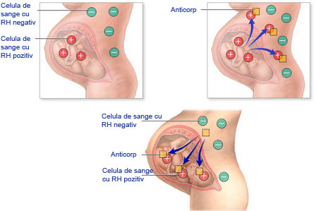
Aplicaţia directă a determinării factorului Rh este diagnosticul prenatal prin amniocenteză , pentru a depista incompatibilitatea feto-maternală. Aceasta incompatibilitate stă la baza bolii prenatale numită eritroblastoză fetală sau boala hemolitică a nou-născutului. Boala apare ca urmare a imunizării mamei cu Rh negativ împotriva antigenului D de pe membrana eritrocitelor fătului cu Rh pozitiv, moştenit de la tată . La naştere hematiile purtând antigenul Rh ajung în sângele mamei iar aceasta va sintetiza anticorpi anti-Rh. La o nouă sarcină (făt cu Rh pozitiv) celulele de memorie vor sintetiza anticorpii anti-Rh care ajung în sângele fătului şi determină apariţia eritroblastozei fetale.
|